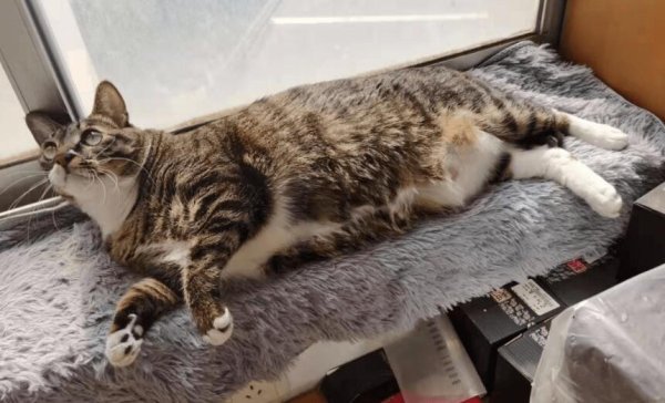
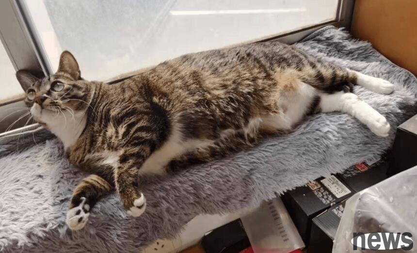
Compulsory course for raising cats: Some basic knowledge about the heart of a cat! The heart is an important functional organ of the body. It transports blood to the whole body and plays a leading role in the internal organs of the body. The same is...
Compulsory course for raising cats: Some basic knowledge about the heart of a cat! The heart is an important functional organ of the body. It transports blood to the whole body and plays a leading role in the internal organs of the body. The same is true for cats. The heart also plays a leading role in the overall situation. The heart is closely related to blood, and blood is the basis for normal activities of the body. Therefore, if you want to understand the cat and understand the material activities in the cat's body, you might as well start by understanding the heart.
1. What are heart rhythm, heart rate and heart sound?
The heart constantly contracts and relaxes, forming rhythmic and regular pulsations. People call this rhythm of pulsation heart rhythm; and the time spent on each contraction of the heart, plus the corresponding diastolic, is called a cardiac cycle. Heart rate refers to the number of heartbeats per minute.
Heart sound is a sound that occurs during the cardiac cycle, the myocardium contracts, the valve opening and closing, and the blood flow rate on the mechanical vibration of the cardiovascular wall. It can be transmitted to the chest wall through surrounding tissue, which can be heard by placing a stethoscope on certain parts of the chest wall.
2. Do you know the position and shape of the heart?
The heart is located in the chest cavity, about 2/3 on the left side of the midline of the body, 1/3 on the right side of the midline, and the front is the sternum; the back is the esophagus, large blood vessels and spine; the sides are lungs, so the heart is strongly protected. The heart is shaped like a crooked pear, with a wide bottom of the heart facing the upper right and a tip facing the lower left. Because the heart is where the large blood vessels enter and exit, it is fixed. The apex can move freely within a certain range.
The heart is covered with two thin and smooth films, called the pericardium. There is a gap between the two layers of pericardial membranes, called the pericardial cavity, which contains a small amount of light yellow to transparent liquid, called pericardial fluid. Pericardial fluid plays a lubrication role when the heart beats, which can reduce friction and resistance. The pericardium is located on the periarthritis and has the function of protecting the heart from excessive expansion.
3. What is the internal structure of the heart?
The convincing appearance seems to be very simple, but its internal structure is relatively complex. If you cut the heart open and remove the blood, you can see that the inside is divided into two parts: the left and right heart by a middle septum. The left heart wall is slightly thicker; the right heart wall is thinner. The left and right hearts are each made of a thin film (called a petal) that looks like a flower, separated into upper and lower halves, with the upper atrium and the lower ventricle. Because the valve is located between the atrium and the ventricle, it is also called the atrioventricular valve. The valve between the left atrium and the left ventricle is composed of two pieces, called the mitral valve. The valve between the right atrium and the right ventricle is composed of three pieces, called the tricuspid valve. The left atrium and right atrium, and the left ventricle and right ventricle are not connected since birth. The atrium receives blood back from the extracardiac blood vessels (systemic veins and pulmonary veins), and then squeezes into the ventricle through the valve, which pumps the blood into the peripheral aorta and pulmonary artery with its powerful contraction force. The valve only allows the atrial blood to flow to the ventricle, and does not allow the ventricle blood to flow back into the atrium. The one-way opening of the valve ensures that the blood flows in a single direction.
4. Why does the heart beat day and night?
Because the heart has a special performance, namely self-discipline. In animal experiments, we can see that even if the heart is taken out of the animal's body, the heart can continue to beat for a certain period of time. Where does this self-discipline of the heart come from? It turns out that there are self-discipline cells that other organs in the body do not have. Self-discipline cells are like small power stations. They can automatically and rhythmically emit tiny currents without any external stimulation or nerve stimulation, stimulating the myocardium to contract and produce beating. This self-regulatory cell of the heart is concentrated at the entrance of the superior vena cava of the right atrium, forming a special nodule called sinoatrial node. The sinus node is like a pulse generator, constantly sending electrical signals, and transmits them downward through a fine conduction system, thereby directing and controlling the heart to beat rhythmically day and night.
5. Some common heart diseases in cats
1. Hypertrophic heart disease
Among cats' heart diseases, the incidence of hypertrophic heart disease can be said to be the highest. Once a cat is diagnosed with this disease, it begins to race against the disease and fight against life for a long time.
Cats with mild lesions may not have any symptoms at the beginning. As the condition develops, cats will experience symptoms of increased breathing frequency, weakness, exercise intolerance, tachycardia, and even difficulty breathing and fainting. Most cats are treated with dyspnea or acute hind limb paralysis. At this time, hypertrophic heart disease has reached moderate or even severe conditions, and the cat may die suddenly. Hypertrophic heart disease may occur when cats tend to become adults, and the high incidence age is young and middle-aged (2 to 8 years old). In pet hospitals, many purebred cats are common confirmed targets of this disease, especially Maine cats, Persian cats, American shorthair cats, Norwegian forest cats, British shorthair cats, Scottish fold-eared cats, etc.
2. Pericardial effusion in cats
Pericardial effusion refers to abnormal accumulation of fluid in the pericardial. Cardiac tamponade refers to excessive pressure in the pericardial, which is often secondary to pericardial effusion, which leads to an increase in the intracardiac pressure, which in turn affects the filling of the ventricle during the diastolic period, reducing the heart's stroke volume and reducing cardiac output. The pericardial effusion in cats is mostly infectious (infectious peritonitis, bacterial infection), tumor, cardiomyopathy, uremia, trauma, left atrium rupture, and a certain idiopathic disease.
The clinical symptoms vary according to the increase rate of pericardial exudate and the degree of heart compression. Mild exudation does not show obvious clinical symptoms, and moderate effusion and slow accumulation can lead to right heart failure. Symptoms are generally:
A. Tiredness, anorexia, atrophy,
B. Difficulty in breathing, rapid breathing, and cough caused by pleural effusion or severe ascites.
C. The abdominal circumference is enlarged due to ascites or hepatomegaly
D. Faint, often overactivity, large amounts of effusion or effusion speed are too fast, often leading to decreased cardiac output. Symptoms generally include:
A. Significant atrophy or paralysis
B. Difficulty in breathing or speeding up
C. Sudden death

6. How does blood circulate in the heart and blood vessels?
According to the different circulation paths of blood in the body, blood circulation can be divided into systemic circulation and pulmonary circulation.
System circulation: When the heart contracts, the fresh blood contained in the left ventricle is first pumped to the aorta and flows through the aorta to the capillaries in various parts of the body through the aorta, transporting oxygen and nutrients to various organs and tissues and cells for material exchange, and taking away the waste and carbon dioxide produced by metabolism, becoming venous blood, and finally collecting it into the upper and lower vena cava and returning to the right atrium. This cycle process is called the system cycle, also known as the large cycle.
Pulmonary circulation: After the venous blood received from the right atrium flows into the right ventricle, it is first injected into the pulmonary artery, and is injected into the pulmonary capillaries through the pulmonary artery, and gas exchange is performed in the lungs, fully absorbing oxygen and expelling carbon dioxide, the blood becomes oxygen-containing bright red blood, and then sent back to the left atrium from the pulmonary vein. This cycle is called pulmonary circulation, also known as small circulation. After the blood circulating through the lung reaches the left atrium, it is squeezed into the left ventricle, and then pumped into the aorta from the left ventricle to deliver to all parts of the body, repeating over and over again. Therefore, the systemic circulation and the lung circulation are connected through the heart, forming a blood circulation together.